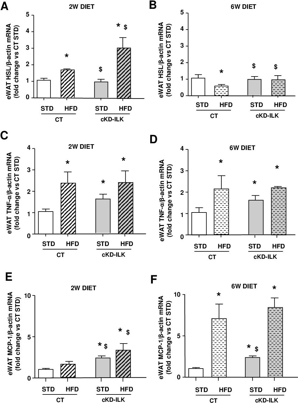
Fig. 5. Adipose expression of lipolysis and inflammatory markers from CT and cKD-ILK during short-term STD or HFD challenges. Fresh isolated epididymal white adipose tissue (eWAT) explants were extracted from CT and cKD-ILK fed with either STD or HFD for 2 or 6 weeks (w). Animals were maintained in fasting conditions before to extract the tissues and proceed to the determinations. Lipase HSL (A, B) and pro-inflamatory adipokines TNFα (C, D) and MCP-1 (E, F) mRNA expression levels fold changes, analyzed by RT-qPCR and normalized to β-actin as endogenous control. N=12 per group. All data are represented as means ± SEMs. *= P<0.05 vs CT STD, $=P<0.05 vs CT HFD.